|
A medical course, like you've never seen before...
Built from scratch, and designed exclusively for iOS, this application takes advantage of what makes the iPad so different...at your benefit :
- mobility
why bother blocking 2 hours for learning behind a PC screen ? Thanks to the tablet, learning has never been so easy. You can do it at your pace, when you are ready, available, focused. And even in places you'd never thought about : plane, train, waiting room...
- interactivity
this app is not a static Powerpoint slideshow. Here is why you will never get bored with it...
- It is non linear: do you want to read the courses before training on cases ? Do you want to read all literature after each case, or do you prefer ground based explanations? Do you want to read cases by diagnosis, topic (e.g: anterior cancers, prostatitis,...), randomly, by progressive difficulty ?
- It tracks your progression: You can follow your overall progression and your score for each case, and watch your progress over time.
- You can compare yourself with others: All cases have a dedicated answering grid showing a statistical representation of the other users answers. This is a great way to compare yourself with your colleagues, and to control your progression.
- You have a direct link with the experts: Each case can be discussed with the team using built-in email button.
- designed BY and FOR radiologists
|
|
| |
|
| Clinical Cases |
|
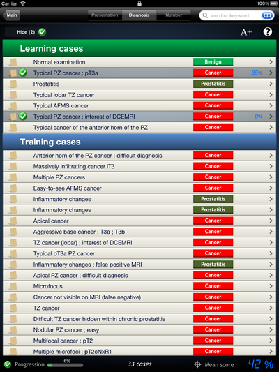
- 33 cases, prepared by specialists in prostate imaging and urologists, taken from real-life activity, regularly and automatically updated when new follow-up events are known...
| - Learning cases (typical presentation) |
7 cases |
| - Training cases (full version) |
26 cases |
| - MRI examination performed before the first series of biopsies (examination-virgin patient) |
21 cases |
| - History of negative biopsies |
12 cases |
| - MRI examination performed at distance of biopsies (without artifact) |
all cases |
| - 12-core randomized TRUS biopsies |
all cases |
| - Negative biopsies |
8 cases |
| - Positive biopsies |
25 cases |
| - Posterior cancers |
19 cases |
| - Anterior cancers |
9 cases |
| - AFMS cancers |
3 cases |
| - Prostatitis or inflammatory changes |
6 cases |
| - Prostatectomy proof |
18 cases |
| - Proven pT3 |
10 cases |
| - Microfoci (true or false) |
8 cases |
- Cases can be sorted by presentation, diagnosis, number, institution
- 4 sections in each case : patient history, images, quiz, discussion. You can switch from one to another in the twinkle of an eye.
|
|
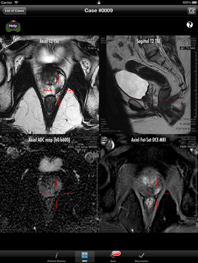
- Images
- Approx. 4000 images
- Immediate access to images (no loading time), no time to waste setting up the series on the screen. All is there. Period.
- Images displayed at a resolution of 320x320 (more than the acquisition resolution)
- All series are described and sometimes commented (technical details)
- Seamless scrolling through synchronized series with up/down swipe
- Real-time reference line on a coronal or sagittal view
- Zoom to full-screen for each series using pinch-in / pinch-out
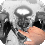
- Touch any part of the image to view a red 3D synchronized pointer
- Hold your finger on a detail to view it zoomed in full screen
- Instant interpretation help wizard in the "learning cases" section
- 1.5 and 3T multiparametric imaging (majority at 1.5T, on a Philips Intera device) using reliable morphologic and functional imaging sequences :T2-w, diffusion-w and DCE-T1w imaging (no spectroscopy). Most of the time in axial oblique reference plane.
|
 |
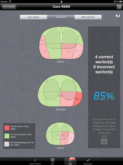
- Standardized 27-region schematic interpretation grid (recommended by the PREDICT and ESUR 2012 guidelines) to...
- Evaluate yourself in comparison with a correction grid (adapted from biopsy and/or prostatectomy findings)
- Send your grid (anonymously) to our servers, populate our answers database and compare your findings with those of other users worldwide (statistical grid of all answers for the case). This feature is HUGE. It allows us select the easiest and hardest cases, too.
|
 |
- Highly detailed discussion section, including "MRI findings", "Biopsy report", "Treatment options", "Radical prostatectomy", "Follow-up", "Conclusion", "Take home messages", "References".
- In "MRI findings", a long description of the most significant images of the examination is made, and suspicious lesions are scored using a 5-points scaled "Likert-like" score, with explained criteria, as recommended by the ESUR 2012 guidelines. This helps you describe the lesions in a more standardized way.
- In "Biopsy report", a detailed report of the 12 biopsies, plus optionally targeted cores is given, providing extensive information on prostate sampling (location of positive cores, length of cancer in each core, Gleason score in each prostate area). Results are visible in the 27-region standardized grid.
- In "Radical prostatectomy", all the details about this intervention are given, plus, in some cases, a schematic drawing of all cancerous images in the gland (after pathology reconstruction). This is a tremendous feature, allowing you to come back on the images and watch how benign and cancerous tissue looks at multiparametric MRI.
- In the "Follow-up" section, we included all significant data obtained after surgery or other treatment : recurrence, PSA undetectable, potency or continency information. This section is regularly updated as patients come back in consultation in our urology department. Automatically.
- In "Conclusion", we discuss the important points of the case: what had to be detected on the images, why you couldn't in certain cases, why the case was important for you.
- The "Take-home messages" summarizes all important notions that were discussed in the case, from a list of messages that will be repeated in other cases, and that you should know by heart at the end of the course.
- The "References" section is a great added value to each case: we selected the papers in relationship with the messages evoked in the case. These papers will illustrate our message differently, sometimes with deep details (e.g: ISUP classification of pT3,....). Each reference is a "link", meaning that depending on the case, you will open the Pubmed link, the open-access PDF, or even the PDF you have loaded in the app or on your Dropbox account. Isn't it GREAT ?
|
|
| |
|
Keys for interpretation |
|
Practicing is obviously the most important, but sometimes, you'll need to sit back and read the fundamentals, take time understanding the basic concepts of prostate imaging.
We (Philippe PUECH, professor of radiology, and Arnauld VILLERS, professor of urology in Lille University Hospital, France) have written a series of 14 small flash cards with the essentials of :
- Morphometric and Histopathological concepts
- Zonal anatomy, Anterior and posterior cancers, cancer origins, multifocality, cancer aggressiveness markers
- Multiparametric MRI
- Indications, imaging sequences, semiology by anatomic zone, MRI limitations, MRI performance for the diagnosis of cancer and for staging, extraprostatic assessment, seminal assessment, nodes assessment, how to perform MRI-targeted prostate biopsies
|
|
| |
|
| References |
|
This section is a mine filled with a compilation of the most important references in the field of our program.
- 88 references, each providing a link to an Internet reference database (NCBI PubMed™ by default) plus, if available, a link to the open-access PDF file.
- Manual import of new references, one by one, or grouped (NCBI PubMed™ Export to XML format)
- Abstract, PMID, authors and citation data available for all articles, without Internet access.
- PDF import through direct Internet access (just like you would do within Safari), meaning that if you have the rights to view the PDF in an Internet browser, then you can import it in the app.
- PDF import through a Dropbox account (put the PDF files in your "Prostate MRI" dropbox folder, and they will be automatically visible in the app)
- Customizable Internet database link (allowing direct access to university proxies, and fulltext access in some cases)
- Printing of HTML pages or of the PDF files using iOS built-in print feature (AirPrint)
- Sending of the PDF files by email (to anybody)
- "Send to..." any other PDF-aware app installed on your iPad (GoodReader™, Adobe Acrobat™,...)
- Proprietary annotations of the PDF files, also visible in the list.
- Backup from the app to your Dropbox account (PDF downloaded within your iPad app can be "pushed" or backuped to your PC/Mac)
- Access to all references from within the clinical cases, meaning that if you have already downloaded a PDF, it will be available anywhere within the app.
|
|
| |
|
| Radiologic anatomy |
|
- 40 high-resolution (512x512) annotated images of a male pelvis
- Easily scroll though the series with finger swipe
- >800 "hot points" pinned on the images
- Touch one to view its details : name (international), type (organ, vessel, structure, space, bone, muscle), comments
- Scrolling through images keeps the structure highlighted, so that you can follow it on the series.
- Customizable display of organs, vessels, anatomical structures, spaces, bones, and muscles
- Specific comments on certain structures
|
|
| |
|
| General |
|
- Designed and tested on iPad 1, iPad 2 and new iPad with identical performances
- Optimized for retina display
- English and French versions available
- Anonymous or personalized mode
- No user-data (except your username) is collected
- iCloud backup enabled
- Can be used offline (all cases are bundled within the app)
- Internet access required for updates, Internet browsing, import of references from Internet, Dropbox connection, viewing others responses.
- Reset of user navigation history and answers
- Contextual help available everywhere (where to click, what you can do with your fingers on the screen, what is expected from you, where to go next, how to interpret the images...)
- Lanscape mode available in all Internet and PDF viewing sections
|
|
|